Japan
1 day
RUSH in Emergency Workshop (Tokyo)
Course highlights
- Rapid Ultrasound in Shock and Hypotension (RUSH)
- Application of eFAST, Aorta, IVC, DVT, Lung & BELS protocols
- Hands-on practical scanning
- Case based discussion
- Clinical integration of findings
Course Dates
Description
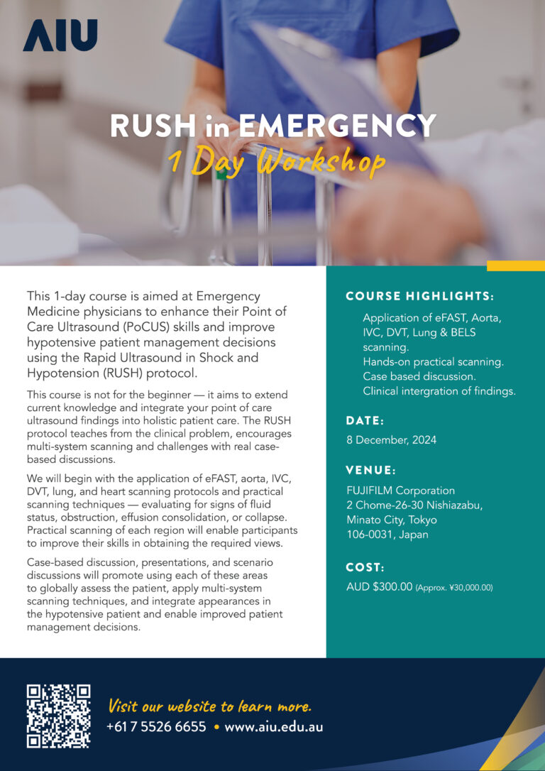
This 1-day course is aimed at Emergency Medicine doctors to enhance their Point of Care Ultrasound (PoCUS) skills and improve hypotensive patient management decisions using the RUSH protocol.
This course is not for the beginner — it aims to extend current knowledge and integrate your point of care ultrasound findings into holistic patient care. The RUSH protocol teaches from the clinical problem, encourages multi-system scanning and challenges with real case-based discussions.
We will begin with the application of eFAST, aorta, IVC, DVT, lung, and heart scanning protocols and practical scanning techniques — evaluating for signs of fluid status, obstruction, effusion consolidation, or collapse.
Practical scanning of each region will enable participants to improve their skills in obtaining the required views.
Case-based discussion, presentations, and scenario discussions will promote using each of these areas to globally assess the patient, apply multi-system scanning techniques, and integrate appearances in the hypotensive patient and enable improved patient management decisions.
Who should attend?
- Emergency Medicine Physician
- Emergency Medicine Consultant
- General Physician
- Internal Medicine Physician
- Internist
Objectives
The learning objectives of this RUSH course are:
- Learn how to utilise multi-system scanning to diagnose and augment clinical decision in the shocked or hypotensive patient.
- Learn how to differentiate causes of shocked stage (cardiogenic, obstructive, distributive, hypovolemic).
- Learn how to recognise signs of shock and fluid status by assessing heart, lungs, aorta, IVC, abdomen, pelvis, and proximal lower limb (DVT).
- You will acquire a complete set of diagnostic quality views of the heart, lung, eFAST, aorta, IVC and DVT to determine shock and hypotensive causes to enable appropriate management decisions:
- Is there free fluid in the chest or abdomen?
- Is there a pericardial effusion?
- Are there signs of cardiac tamponade?
- Is the relative size of the chambers normal?
- Is the LV/RV systolic function preserved?
- Is there any sign of massive pulmonary embolism?
- Is there any sign of hyperdynamic status/hypovolemia?
- Is lung sliding present?
- Is there a pneumothorax?
- Is there any sign of pulmonary oedema?
- Is the pleura normal?
- Is peripheral pulmonary consolidation is present?
- Is there a pleural effusion, if so what size and nature?
- Is there an AAA?
- Is the IVC collapsing?
- Is there a DVT ?
Brochure Download
Click the link below to download the course brochure.
Outcomes
The expected learning outcomes for this course are:
- Utilise multi-system scanning to diagnose and augment clinical decision making e.g. cause of shocked stage (cardiogenic, obstructive, distributive, hypovolemic).
- Recognise signs of shock and fluid status by assessing heart, lung, aorta, IVC, abdomen, pelvis, and proximal lower limb (DVT);
- Integrate ultrasound findings of the shocked patient to affect patient management decisions.
- Identify the parameters of safe application of ultrasound and its’ limitations in medical diagnosis in assessing eFAST, lung, heart, and DVT in proximal lower limb.
- Demonstrate effective use of an ultrasound machine and optimise an ultrasound image.
- Recognise normal ultrasound anatomy and describe it using sonographic terminology.
- Identify the limitations of ultrasound and determine when to on-refer patients for formal scanning or complimentary investigations.
- Acquire images of heart (Sub Xiphoid, PLAX, PSAX, 4C), lung (12 point protocol), abdomen (RUQ, LUQ, pelvis), aorta, IVC, and proximal lower limb (DVT).

HOURS
| Theoretical | 2.75 |
| Practical | 4.25 |
| Total | 7 |

VENUE
FUJIFILM Corporation
富士フィルム
2 Chome-26-30 Nishiazabu,
Minato City, Tokyo
106-0031, Japan

CATERING
Morning tea, a light lunch and refreshments are provided each day.